Pinguecula & Pterygium Treatment
Pterygium and Pinguecula are one of the oldest pathologies known to eye surgeons. Surgery for this condition can range from simple excision to techniques with exotic detail and meticulous maneuvers with task-specific instruments beckoning an era of raised expectations and cosmetic outcomes in the field of ocular surface surgery itself.
Pterygium
Pterygium is a pinkish-yellow, triangular-shaped tissue growth starting from the nasal area of your eye and grows towards the cornea (front, clear window of the eye). This lesion can be varied in its appearance from small and pink to large and angry red with symptoms of dry eye, cosmetically unacceptable appearance and/or adversely affecting vision.
Pinguecula
Pinguecula are yellowish, slightly raised lesions that form on the surface tissue of the white part of your eye (sclera) close to the edge of the cornea. They are typically found in the open space between your eyelids.
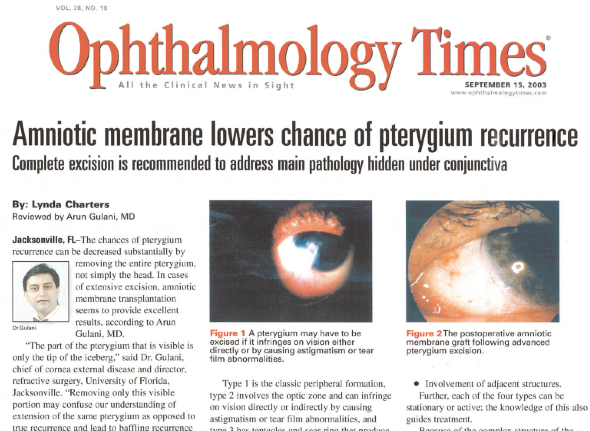
Dr. Gulani performs a specialized surgery for Pterygium and Pinguecula wherein he executes a complete removal of this lesion from it's roots (Ice Berg concept) followed by an Amniotic membrane (Human Placenta) graft and a drug called Mitomycin C. Dr. Gulani uses no stitches during this surgery and instead applies a special glue to hold this graft in place.
Many patients with Pterygium and Pingueculas also have associated vision issues for which they may be wearing contact lenses. Their associated nearsightedness (myopia), Farsightedness (Hyperopia) and Astigmatism can be corrected with Advanced Lasik surgery.
These patients may also have associated Dry eyes which can be corrected by Dr.Gulani's advanced dry eye treatments to eventually help such patients have successful amniotic surgery and also vision surgeries like Lasik and cataract surgery.
Patients from all over the world have come to Gulani Vision Institute to seek Dr. Gulani's skill in this field and many have been pleasantly surprised at the excellent cosmetic appearance of their eyes. Though complications from any surgery are possible as are recurrences, Dr. Gulani's outcomes have raised the bar for these surgeries in also seeking a goal of life without glasses and contact lenses in such cases. The concept of Look Good, See Good!
Having classified Pterygia into 4 categories previously, Dr. Gulani has continuously studied the presentations and outcomes in improving the surgical approach and outcomes over the years to add an additional way to classify the pterygium based on the adhesion of the head to the ocular surface and vascularity along with the draw test on the cornea. Pterygiums can therefore have:
- Head/Neck Adhesion: Peripheral / Central adhesion (this can further be classified into diffuse and focal) In cases of peripheral adhesion the pterygium easily peels off the cornea
- Vascularity: Engorged, tortous vessels and simultaneous conjunctival fold contracture signifies a more aggressive pterygium. This same concept can be used to determine outcomes postoperatively
- Draw test: On tugging on the cornea some pterygia may be small in size but outright gritty and deep into the cornea resulting in thin cornea when removed. Preparation for this before surgery helps plan a smooth outcome (Amniotic graft itself can be used as a lamellar fill). Also removal of these pterygia is more difficult from the corneal surface.
 Thus the basis of Dr. Gulani's surgical steps involve a lamellar approach along with atraumatic pterygium, pinguecula removal as a conjunctival scar with full dissection right up to the roots , including all arising heads followed by subconjunctival mitomycin-C application and Amniotic graft layering on the sclera with glue and reconstructing the fornix in many cases.
- Criteria for surgical plan:
- The extent of the pterygium
- Density of the pterygium
- Involvement of adjacent structures
- Draw test
- Head/Neck adhesion
- Vascularity
Dr. Gulani strives to keep raising the bar on safety and outcomes in this surgery and all patients are made to walk up to a mirror next day (One day post-op) to appreciate look at their operated eye. This "No stitch, No patch and No red" technique along with absence of visual deficit is raising the bar in patients now seeking this approach for related ocular surface conditions like conjunctivochalasis and pinguecula. Here again, since the bar has been raised on outcomes, pinguecula removal follows the same principle as pterygium surgery.
Thanks to Dr.Gulani's perseverance in raising the bar, Ocular surface surgeries have reached a status of cosmetic outcomes and will lead into improving many borderline cases for ocular surface suitability for LASIK laser vision correction thus becoming an integral part of every refractive surgery practice (Corneoplastique[TM])

 About Dr. Gulani
Dr. Gulani is a world renowned eye surgeon and Pinguecula Treatment specialist. Former Chief of the Cornea service and Asst. Professor at University of Florida, School of Medicine; he is Founding Director of the Gulani Vision Institute.
Browse Our Site
